Prof. Ch. Gerber em.


Christoph Gerber is a titular professor em. at the Department of Physics, University of Basel, Switzerland. He was a founding member and Director for Scientific Communication of the NCCR (National Center of Competence in Research Nanoscale Science) and acted more than 15 years as a Vice-Director on the board of the SNI (Swiss Nanoscience Institute) at the Univ. of Basel. He was formerly a Research Staff Member in Nanoscale Science at the IBM Research Laboratory in Rueschlikon, Switzerland, and has served as a project leader in various programs of the Swiss National Science Foundation and in the European Framework 6
Achievements
For the past 45 years, his research has been focused on Nanoscale Science. He is a pioneer in Scanning Probe Microscopy, and he made major contributions to the invention of the Scanning Tunneling Microscope and the Atomic Force Microscope (AFM), he is also a co-inventor of Biochemical sensors based on AFM Technology.
He is the author and co-author of more than 180 scientific papers that have appeared in peer-reviewed journals and has been cited more than 50’900 times in ISI Clarivate Web of Science and 74‘500 times (https://scholar.google.com/citations?user=w9b8kbgAAAAJ&hl=de) in cross-disciplinary fields. He belongs to the one hundred worldwide most cited researchers in Physical Sciences. He has given numerous plenary and invited talks at international conferences.
His work has been recognized with multiple honorary degrees and various awards and appeared in numerous articles in daily press and TV coverage. He is co-recipient of the 2016 Kavli Prize in Nanoscience for the Atomic Force Microscope. In 2023 he received the World Cultural Council Albert Einstein Award for Science. In 2024, he was given the status of a Citation Laureate by the Institute for Scientific Information (ISI) at Clarivate. He is a Fellow of the American Physical Society, a Fellow of the World Technology Network and a Fellow of the IOP Institute of physics UK. He serves in the advisory board of several nano institutes and has chaired and co-chaired various international conferences. His IP portfolio contains 37 patents and patent publications. His private interest range from literature (scientific and a good novel) to art and sports (he is a passionate skier and plays an acceptable round of golf).
His current interests include
- Biochemical sensors based on AFM Technology
- Chemical surface identification on the nanometer scale with AFM
- Nanomechanics, nanorobotics, and molecular devices at the ultimate limits of measurement and fabrication
- Atomic Force microscopy research on insulators
-
- Self-organization and self-assembly at the nanometer scale
SPM History
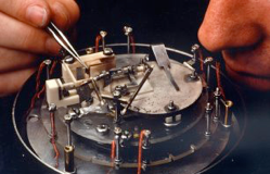
First STM (1981)
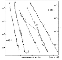
Exponential tunneling current
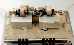
First AFM (1986)
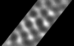
First atomic resolution on graphite
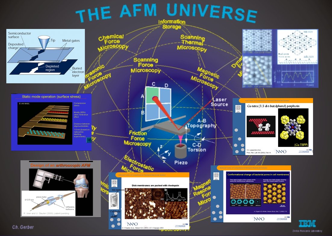
AFM universe
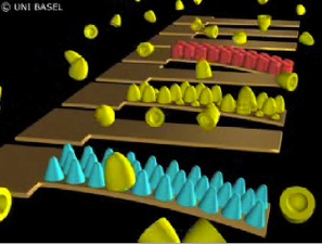
Nanomechanical Sensing with Cantilever Arrays
Cantilever bending
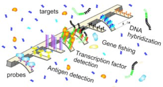
Multifunctional cantilever array sensors
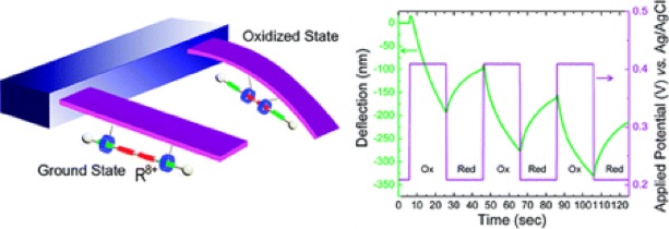
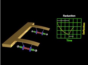
Molecules can drive a nanomechanical cantilever
(related to the work of Fraser Stoddard, Nobel Laureate in Chemistry 2016).
F. Stoddard & P. Weiss groups, ACS Nano 3, 291 (2009).
A Mechanical Actuator Driven Electrochemically by Artificial Molecular Muscles
Oxidation and reduction of bistable rotaxene molecules controlled by electrochemistry produce forces that bend a nanomechanical cantilever.
40 years of Atomic Force Microscopy (AFM), a brief history
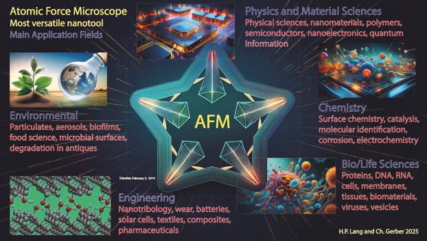
Over the past four decades, Atomic Force Microscopy (AFM) has evolved from a novel imaging technique into one of the most powerful and versatile tools in nanoscience and nanotechnology. By enabling the visualization of non-conductive materials with true molecular and atomic resolution, AFM stepped beyond a shortcoming of the STM and opened the door to an exceptionally broad range of scientific fields – physics, materials science, engineering, chemistry, biology, medicine and beyond, establishing AFM as an indispensable nanotool.
AFM’s central strength lies in its operational principle: a sharp tip, integrated in a microcantilever, measures forces interacting with a surface with extraordinary sensitivity, converting these minute interactions into topographic maps with unprecedented resolution. Researchers can study samples in ultrahigh vacuum, ambient conditions, liquids, controlled atmosphere, at any temperature level making AFM also uniquely suited for biological and soft-matter studies. This adaptability drove early enthusiasm, and continuous technological refinements transformed AFM from a delicate research prototype into a routine analytical instrument.
A major driver of AFM’s rise has been its expanding modes of operation. AFM now measures a whole conglomerate of atomic forces, e.g mechanical, electrostatic , van der Waals, magnetic, friction and chemical interactions at the nanoscale. Techniques such as contact mode, non-contact dynamic mode (tuning fork), tapping mode, Kelvin force mode, peak-force and off- resonance wave mode, quantitative nanomechanical mapping have effectively turned AFM into a multifunctional toolbox rather than a single instrument. Each new mode extended AFM’s reach into previously inaccessible scientific questions, reinforcing its reputation as a platform rather than a static technology.
Another reason for AFM’s enduring power is its synergistic integration with other methods. Correlative AFM–optical microscopy, AFM–Raman spectroscopy, SEM Scanning electron microscopy, HIM helium ion microscopy and other methods have enabled studies in a plethora of scientific processes at the nanoscale and thus provide more comprehensive data than a single technique can. These systems have kept AFM at the forefront of nanoscience, even since competing techniques such as super-resolution optical microscopy and advanced electron microscopy have advanced dramatically.
In biology AFM’s single-molecular spectroscopy revolutionized biological and materials science by allowing direct manipulation and measurement of individual molecules, providing insights into their mechanical properties, folding pathways, and interactions. High speed AFM opened up the time domain of chemical activity by monitoring the cellular machinery at the nanoscale with milliseconds resolution. Ultra-rapid and highly sensitive nanomechanical cantilever array sensors impacted medical diagnostics by surpassing time-consuming current gold standards.
Today, forty years after its invention, AFM remains unmatched in its combination of spatial resolution, environmental versatility, and multifunctionality. Its ability to probe surfaces physically, electrically, biologically and chemically—often simultaneously—has made it the cornerstone of nanoscale characterization. Far from being surpassed, AFM continues to innovate, proving that its fundamental principle is robust, adaptable, and with the integration of machine learning and AI remarkably future-proof.